Research
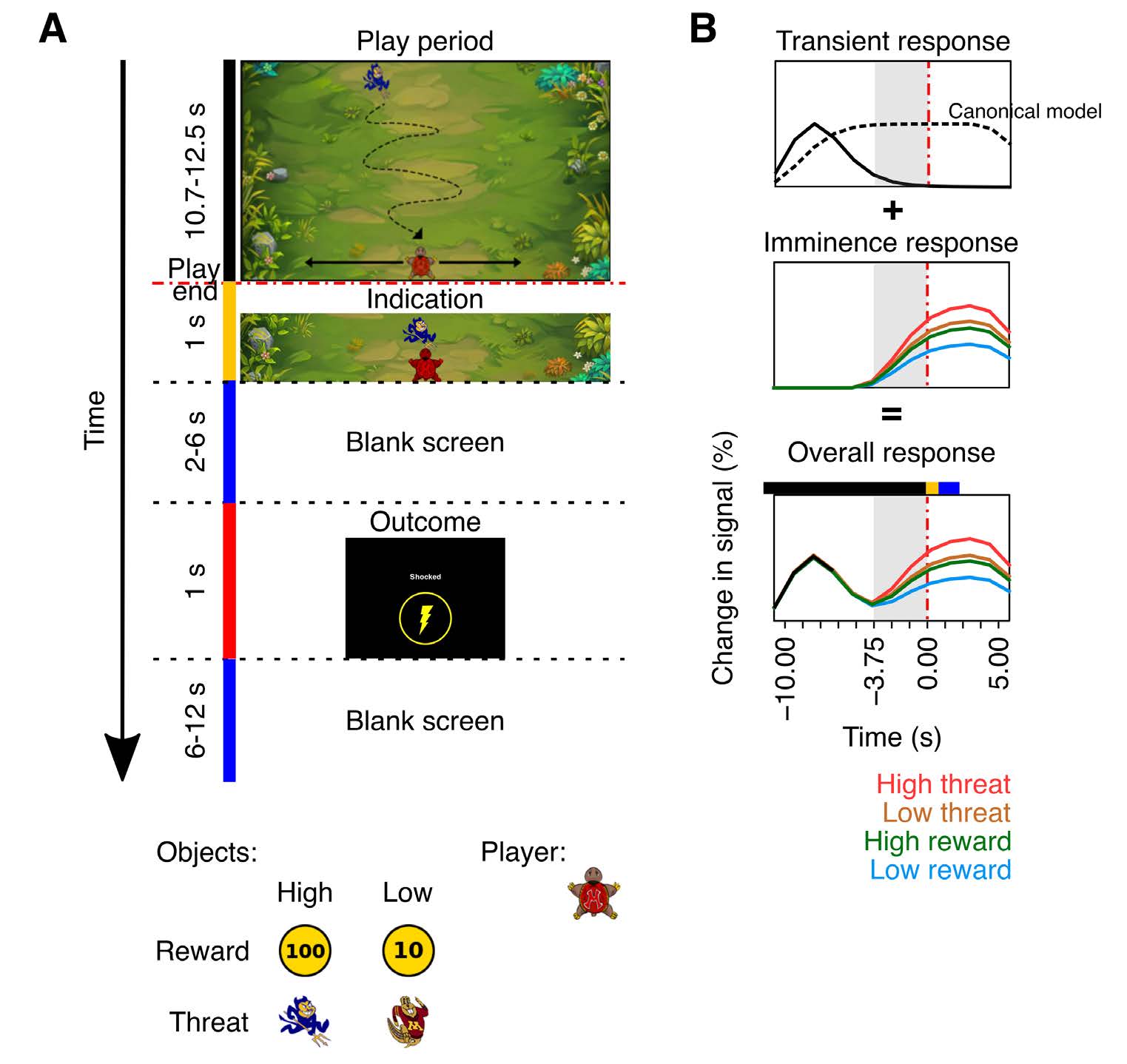
Threat and reward imminence processing in the human brain
Dynamics of aversive and appetitive processing while engaged in comparable trials involving threat-avoidance or reward-seeking
Dinavahi Murty, Songtao Song, Srinivas Govinda, and Luiz Pessoa. bioRxiv, January 2023.
Show More
Abstract
In the human brain, aversive and appetitive processing have been studied with controlled stimuli in rather static settings. In addition, the extent to which aversive- and appetitive-related processing engage distinct or overlapping circuits remains poorly understood. Here, we sought to investigate the dynamics of aversive and appetitive processing while male and female participants engaged in comparable trials involving threat-avoidance or reward-seeking. A central goal was to characterize the temporal evolution of responses during periods of threat or reward imminence. For example, in the aversive domain, we predicted that the bed nucleus of the stria terminalis (BST), but not the amygdala, would exhibit anticipatory responses given the role of the former in anxious apprehension. We also predicted that the periaqueductal gray (PAG) would exhibit threat-proximity responses based on its involvement in proximal-threat processes, and that the ventral striatum would exhibit threat-imminence responses given its role in threat escape in rodents. Overall, we uncovered imminence-related temporally increasing (“ramping”) responses in multiple brain regions, including the BST, PAG, and ventral striatum, subcortically, and dorsal anterior insula and anterior midcingulate, cortically. Whereas the ventral striatum generated anticipatory responses in the proximity of reward as expected, it also exhibited threat-related imminence responses. In fact, across multiple brain regions, we observed a main effect of arousal. In other words, we uncovered extensive temporally-evolving, imminence-related processing in both the aversive and appetitive domain, suggesting that distributed brain circuits are dynamically engaged during the processing of biologically relevant information irrespective of valence, findings further supported by network analysis.
Related Papers
-
Murty, D., Song, S., Surampudi, S., & Pessoa, L. (2023). Biorxiv.
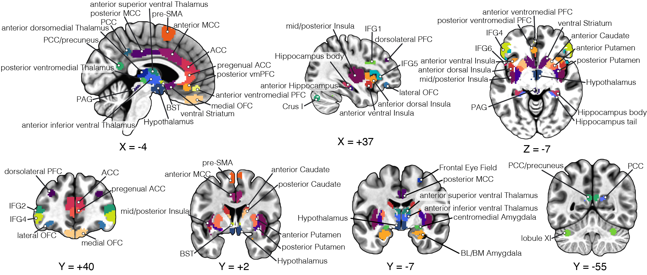
Distributed and multifaceted effects of threat and safety
How anxious apprehension is processed in the human brain
Murty D., Song S., Morrow, K., Kim, J., Hu, K., Pessoa, L. (2022). J Cogn Neurosci.
Show More
Abstract
In the present fMRI study, we examined how anxious apprehension is processed in the human brain. A central goal of the study was to test the prediction that a subset of brain regions would exhibit sustained response profiles during threat periods, including the anterior insula, a region implicated in anxiety disorders. A second important goal was to evaluate the responses in the amygdala and the bed nucleus of the stria terminals, regions that have been suggested to be involved in more transient and sustained threat, respectively. A total of 109 participants performed an experiment in which they encountered “threat” or “safe” trials lasting approximately 16 sec. During the former, they experienced zero to three highly unpleasant electrical stimulations, whereas in the latter, they experienced zero to three benign electrical stimulations (not perceived as unpleasant). The timing of the stimulation during trials was randomized, and as some trials contained no stimulation, stimulation delivery was uncertain. We contrasted responses during threat and safe trials that did not contain electrical stimulation, but only the potential that unpleasant (threat) or benign (safe) stimulation could occur. We employed Bayesian multilevel analysis to contrast responses to threat and safe trials in 85 brain regions implicated in threat processing. Our results revealed that the effect of anxious apprehension is distributed across the brain and that the temporal evolution of the responses is quite varied, including more transient and more sustained profiles, as well as signal increases and decreases with threat.
Related Papers
-
Murty DVPS, Song S, Morrow K, Kim J, Hu K, Pessoa L. . J Cogn Neurosci. 2022 Feb.
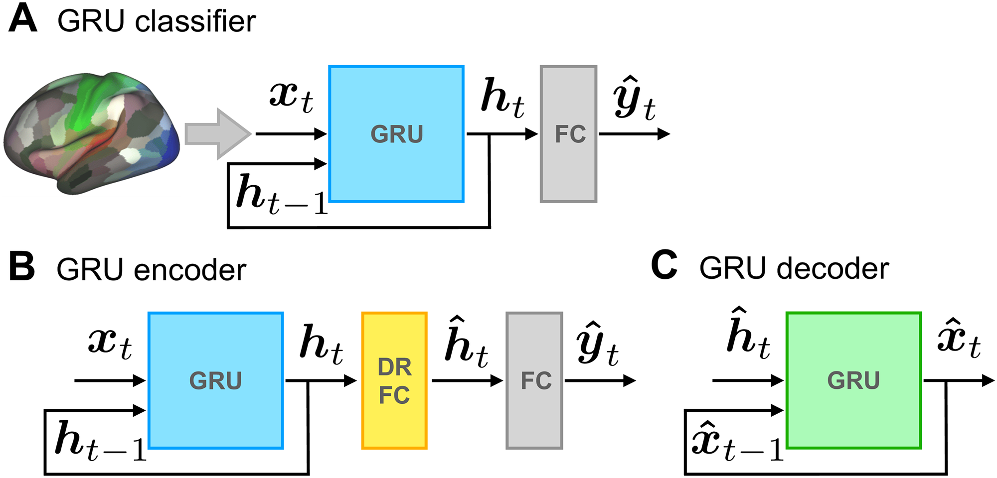
Learning brain dynamics for decoding and predicting individual differences
Brain signals are dynamic and evolve in both space and time.
Misra J, Surampudi SG, Venkatesh M, Limbachia C, Jaja J, et al. (2021) PLOS Computational Biology.
Show More
Abstract
Insights from functional Magnetic Resonance Imaging (fMRI), as well as recordings of large numbers of neurons, reveal that many cognitive, emotional, and motor functions depend on the multivariate interactions of brain signals. To decode brain dynamics, we propose an architecture based on recurrent neural networks to uncover distributed spatiotemporal signatures. We demonstrate the potential of the approach using human fMRI data during movie-watching data and a continuous experimental paradigm. The model was able to learn spatiotemporal patterns that supported 15-way movie-clip classification (∼90%) at the level of brain regions, and binary classification of experimental conditions (∼60%) at the level of voxels. The model was also able to learn individual differences in measures of fluid intelligence and verbal IQ at levels comparable to that of existing techniques. We propose a dimensionality reduction approach that uncovers low-dimensional trajectories and captures essential informational (i.e., classification related) properties of brain dynamics. Finally, saliency maps and lesion analysis were employed to characterize brain-region/voxel importance, and uncovered how dynamic but consistent changes in fMRI activation influenced decoding performance. When applied at the level of voxels, our framework implements a dynamic version of multivariate pattern analysis. Our approach provides a framework for visualizing, analyzing, and discovering dynamic spatially distributed brain representations during naturalistic conditions.
Related Papers
- Misra J, Surampudi SG, Venkatesh M, Limbachia C, Jaja J, et al. (2021) PLOS Computational Biology
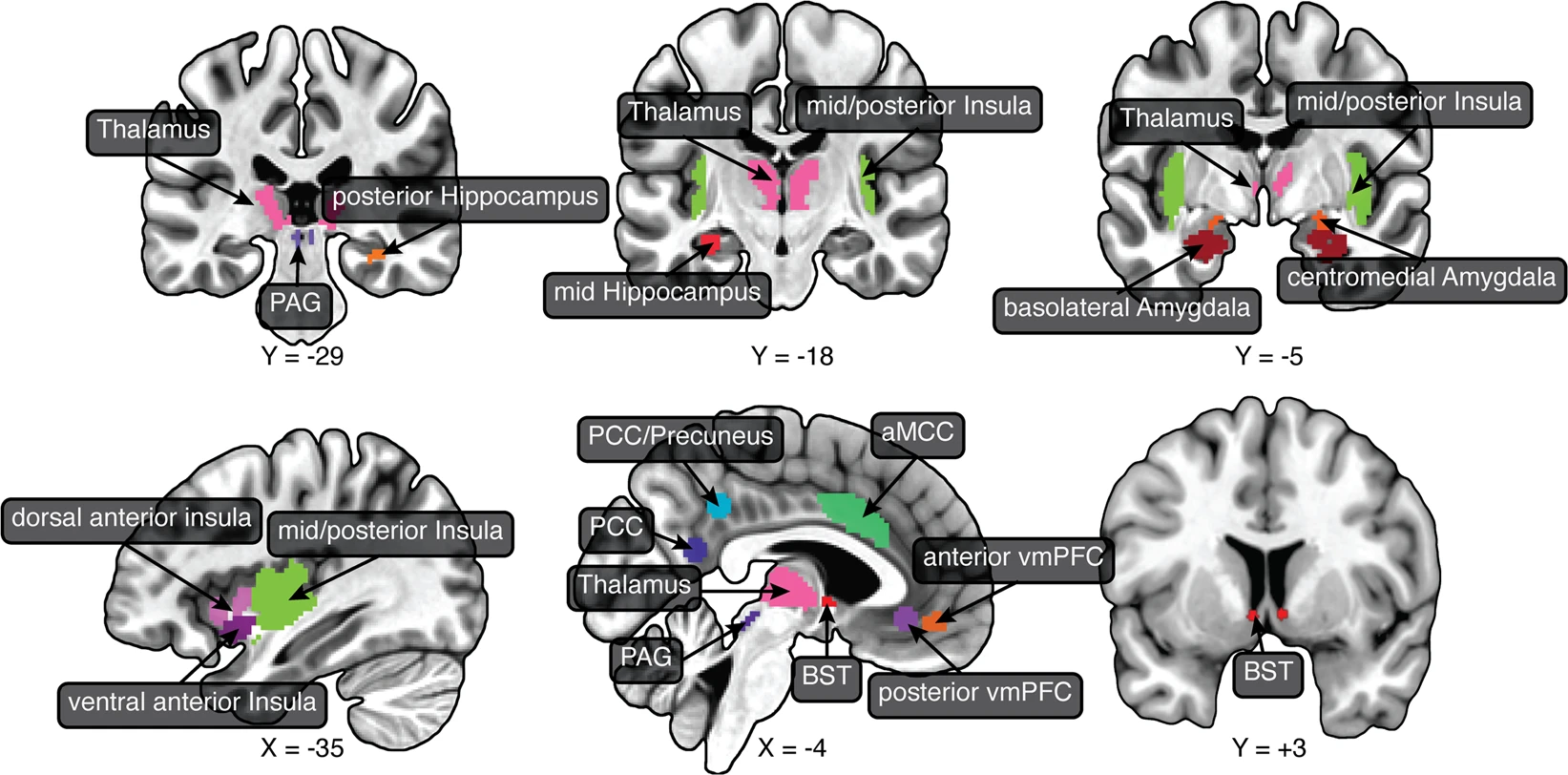
Controllability over stressor decreases response in key threat-related brain areas
The effect of controllability on responses to stressors.
Limbachia, C., Morrow, K., Khibovska, A. et al. (2021) Commun Biol.
Show More
Abstract
Controllability over stressors has major impacts on brain and behavior. In humans, however, the effect of controllability on responses to stressors is poorly understood. Using functional magnetic resonance imaging (fMRI), we investigated how controllability altered responses to a shock-plus-sound stressor with a between-group yoked design, where participants in controllable and uncontrollable groups experienced matched stressor exposure. Employing Bayesian multilevel analysis at the level of regions of interest and voxels in the insula, and standard voxelwise analysis, we found that controllability decreased stressor-related responses across threat-related regions, notably in the bed nucleus of the stria terminalis and anterior insula. Posterior cingulate cortex, posterior insula, and possibly medial frontal gyrus showed increased responses during control over stressor. Our findings support the idea that the aversiveness of stressors is reduced when controllable, leading to decreased responses across key regions involved in anxiety-related processing, even at the level of the extended amygdala.
Related Papers
- Limbachia, C., Morrow, K., Khibovska, A. et al. Controllability over stressor decreases responses in key threat-related brain areas. Commun Biol
Network-level analysis of emotional and motivational processing
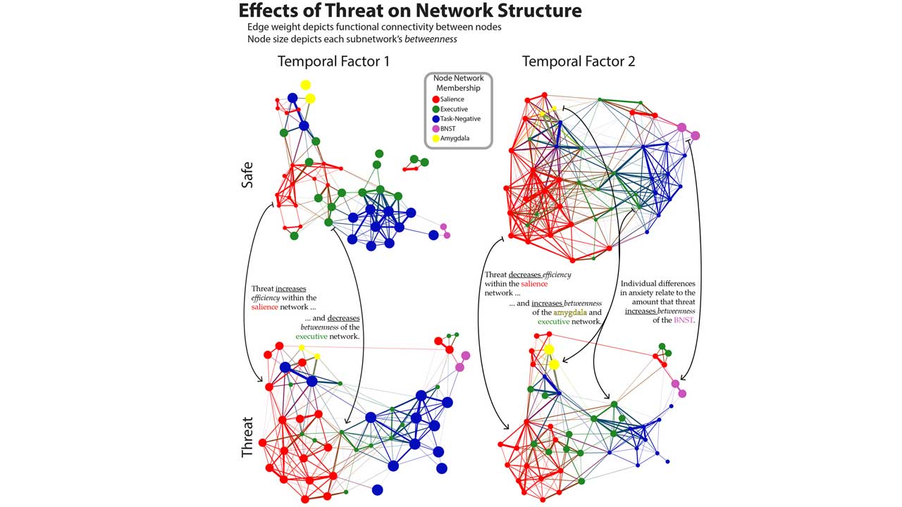
Network organization unfolds over time during periods of anxious anticipation
Brenton McMenamin, Sandra Langeslag, Mihai Sirbu, Srikanth Padmala, and Luiz Pessoa. The Journal of Neuroscience, August 2014.
Show More
Abstract
Entering a state of anxious anticipation triggers widespread changes across large-scale networks in the brain. The temporal aspects of this transition into an anxious state are poorly understood. To address this question, an instructed threat of shock paradigm was used while recording functional MRI in humans to measure how activation and functional connectivity change over time across the salience, executive, and task-negative networks and how they interact with key regions implicated in emotional processing; the amygdala and bed nucleus of the stria terminalis (BNST). Transitions into threat blocks were associated with transient responses in regions of the salience network and sustained responses in a putative BNST site, among others. Multivariate network measures of communication were computed, revealing changes to network organization during transient and sustained periods of threat, too. For example, the salience network exhibited a transient increase in network efficiency followed by a period of sustained decreased efficiency. The amygdala became more central to network function (as assessed via betweenness centrality) during threat across all participants, and the extent to which the BNST became more central during threat depended on self-reported anxiety. Together, our study unraveled a progression of responses and network-level changes due to sustained threat. In particular, our results reveal how network organization unfolds with time during periods of anxious anticipation.
Related Papers
- Joshua Kinnison, Srikanth Padmala, Jong-Moon Choi, and Luiz Pessoa. Network analysis reveals increased integration during emotional and motivational processing. The Journal of Neuroscience. 2012 June 13; 32(24):8361-72.
- Brenton McMenamin and Luiz Pessoa. Discovering networks altered by potential threat (“anxiety”) using quadratic discriminant analysis. Neuroimage. 2015 Aug; 116:1-9.
Mission Statement
Our long-range goal is to contribute to a better understanding of the basic mechanisms by which emotional/motivational and cognitive brain systems interact in the generation of complex behavior. As a step forward to this goal, we are currently investigating how tasks that require “top-down control” and emotional/motivational processing interact in the brain. Cognitive and emotional/motivational systems have been largely considered to function separately from one another. However, a deeper understanding of the brain function and associated behavior requires studying interactions between these systems. Recent techniques, including functional neuroimaging (such as fMRI), allow the study of several brain systems simultaneously and pave the way for the study of interactions in the brain. By studying cognitive-emotional interactions we expect to make contributions to understanding the neural basis of behavior. At the same time, we expect our work to have clinical implications. For example, by providing a better understanding of cognitive-emotional interactions during normal behavior, our research can help understand the mechanisms that potentially go awry in many debilitating mental illnesses.
